A microscope opens a door into a fascinating—and educational—world
Redwoods don’t actually have needles, they have leaves. We call their leaves needles because they’re thin and pointy, but they are indeed leaves, performing all the necessary leafly duties. As it turns out, redwoods even have two distinct kinds of leaves. One kind specializes in making the tree’s food through photosynthesis and the other focuses on absorbing water. It’s a botanically harmonious division of labor.
One of the researchers who figured this out is Dr. Alana Chin, an associate professor of plant physiology at Cal Poly Humboldt. Dr. Chin is part of the university’s Tree Function Lab, where she studies how nimble the redwoods and their pointy little leaves can be when adapting to a warmer, drier climate. (Save the Redwoods League helps fund the Tree Function Lab.)
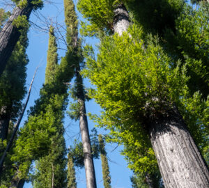
For instance, redwoods along California’s rainy North Coast arrange their leaves so that the ones that absorb water grow low on their trunks, with the photosynthesizing leaves higher up near the tree’s crown, in order to better collect light from the sporadically shining sun. Redwoods further south, in sunnier, drier areas, do the opposite: Their water-absorbing leaves grow nice and high where they can more easily capture fog and rain.
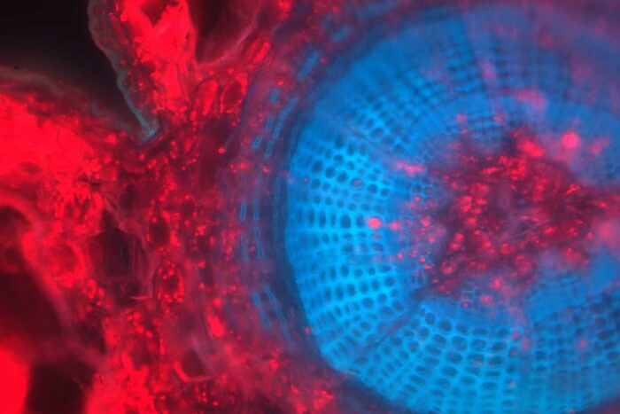
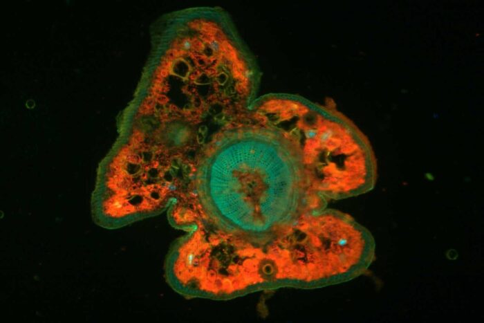
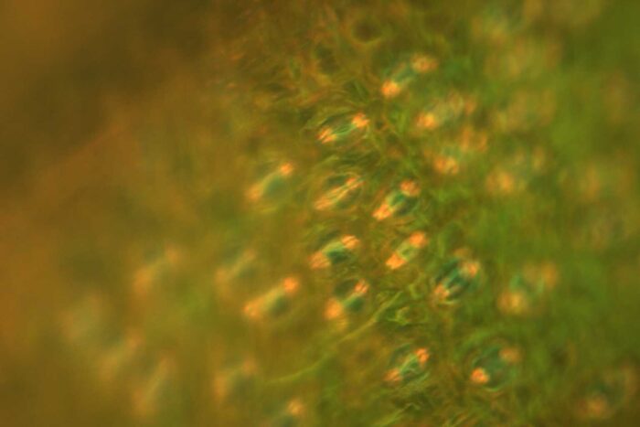
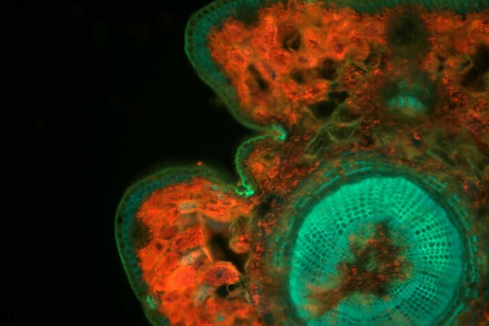
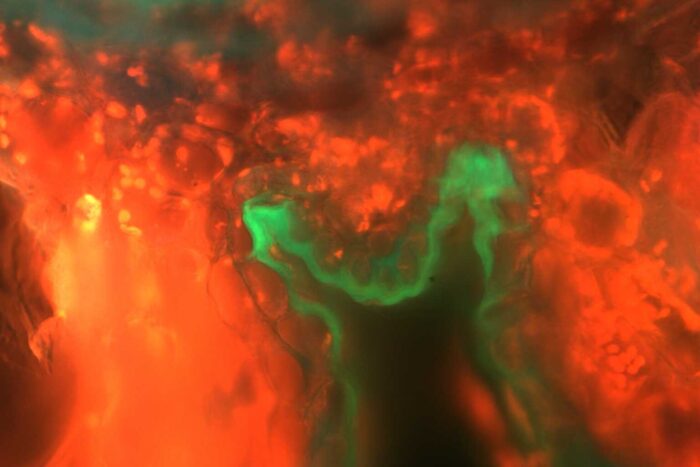
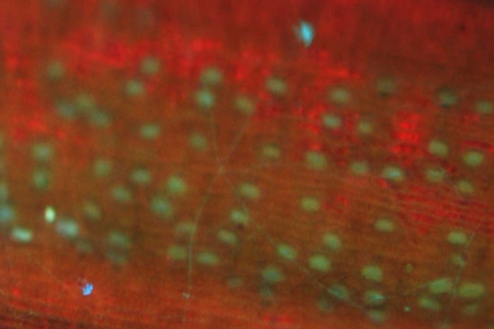
As part of her work, Dr. Chin collects redwood leaves, bathes them in ultraviolet light, and looks at them under powerful microscopes. The resulting images can be stunning. Some look like little nebulae awash in neon colors produced by natural chemicals in the leaves that react to the UV light. Others show highways of brightly glowing stomata—the tiny pores on leaves that allow a plant to breathe.
Dr. Chin analyzes the colors and the arrangement of the stomata to better understand how redwoods absorb water from fog. Eventually, images like these may help researchers predict how redwoods will fare in drier conditions. They may also help inform restoration work by showing us which redwoods will grow healthy and strong in the future.
Plus, the photographs are just plain cool. In the hand, a redwood’s leaf is just a thin strip of green. But there’s an entire universe in that leaf.
This feature appears in the beautiful printed edition of Redwoods magazine, a showcase of redwoods conservation stories, breathtaking photos, and ways you can help the forest. Only a selection of these stories are available online.
Join our thousands of members today for only $25, and you’ll get future editions of Redwoods magazine.


One Response to “Redwood leaves like you’ve never seen them before”
Chris Reå (ISA 185787)
Hi there – well i look after two young redwood trees erroneously planted in pH 5.6^ c.1850 and I have recently discovered evidence for a bright orange fungus growing at the main ‘base anchor’ root flare. a metre and a half above this zone is a continuing black exudate – recent culturing analysis by David McKellar at the Forest Pathology lab. here (Alice Holt Lodge? has now identified
Leptosphaerulina in the slime flux..in addition I have instigated an ultrasound tomography survey that has revealed significant vascular decay within this target area and elsewhere around the base – id there any further reference material useful for CPD (continuing professional development), to guide the works programme in situ (I recommended soil improvement basics back in early December and still there has been no action from mid-management) !! looking forward to further comms. & yes I have climbed this specimen (a small specimen @ 508 cm girth and approx. 40 m. tall, in addition to its sister tree that also has the same developing chlorosis) – both trees are located within a College Campus in the North Oxford (Uk) suburb conservation area and I can confirm that the two Austrian Black Pine trees in situ and a developing veteran Yew tree are also not looking too good in the upper canopy zones – note that a resistograph of the Austrian Pines revealed nil vascular necrosis – however all of the tree items mentioned were subjected to major deadwood removal before last fall etc – last year I travelled to the Napoleon III gardens in Vichy, France to look closer at a similar girth Redwood – also planted to commemorate the Crimea Campaign – although the tree was enclosed by railings, the same chlorosis can be observed and it was disheartening to observe that risk mitigation measures may have compromised the longevity of the plant main stem etc – I have to say tho’ that the general standard of tree care is exemplary – looking forward to further comms., Best regards & thank you for the continuing inspiration